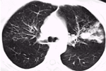
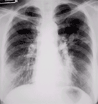
CASE OF THE MONTH

2002

MAR


Fig 1

Figure 1

Fig 2

Figure 2


History

Asthma with repeated hospital admission.
Figure 1 Chest Radiograph
Fingers-in-glove shadows in the left upper and middle zones suggesting bronchocoeles. Reticular shadows in the right upper and middle zones.
Figure 2 High resolution CT scan of lungs
Left upper lobe bronchocoeles are seen. Right upper lobe fibrosis is seen.
 

DIAGNOSIS :

Allergic bronchopulmonary aspergillosis.

HISTORY

PREVIOUS CASES

HOME

COMMENTS

Click on thumbnail for full-sized image


Disclaimer: The material used in this Web page is for educational purpose only.