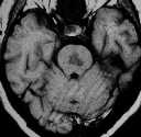
Figure 1
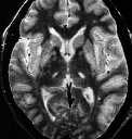
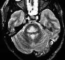
Figure 2
Figure 1 & 2 MR shows a large T1 hypo & T2 hyperintense lesion in the central pon.Figure 3 There are also T2 hyperintense lesions in the bilateral basal ganglia and thalami.
| ||||
HISTORY |
PREVIOUS MTHS |
|||
HOME |
CURRENT MTH |
|||
CASE OF THE MONTH |
|
|
Figure 1 |
Figure 2
|
| ||||||||||
|---|---|---|---|---|---|---|---|---|---|---|---|---|
Click on thumbnail for full-sized image
Disclaimer: The material used in this Web page is for educational purpose only.