Figure 1
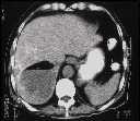
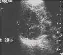
Figure 2
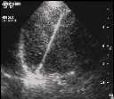
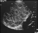
Figure 1 US left lobe of liver showed reticulations.Figure 2 US right lobe of liver showed presence of a large liver abscess.Figure 3 Plain CT showed reticular calcifications & air containing fluid collection in R lobe of liver.Figure 4 Percutaenous drainage of liver abscess under US guidance. 150 ml greenish pus was drained. Culture yielded Clostridium.
| ||||
HISTORY |
PREVIOUS MTHS |
|||
HOME |
CURRENT MTH |
|||