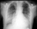
Figure 1
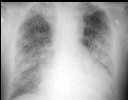
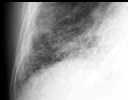
Figure 2
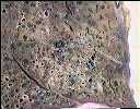
Figure 1 & 2 CXR showed bilateral lower zone reticulo-nodular shadows with honeycomb appearance.Figure 3 CXR showed increase shadows in mid zones.Figure 4 Post-mortem finding of interstitial fibrosis in right middle and both lower lobes. Areas of consolidation in upper lobes with histological changes of bronchiolitis obliterans with organising pneumonia (BOOP)
| ||||
HISTORY |
PREVIOUS MTHS |
|||
HOME |
CURRENT MTH |
|||