
Figure 1
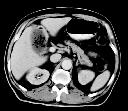
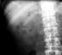
Figure 2
Figure 1 AXR shows collection of gas with the configuration of gall bladder in the right upper quadrant.Figure 2 AXR(RUQ, 36 hours later) demonstrates that there is now mottled and curvilinear gas lucencies in the gall bladder region.There is also gas in the cystic duct.Figure 3 CECT abdomen shows that there is gas in the gall bladder lumen and in the thickened gall bladder wall chacteristic of emphysematous cholecystitis.
| ||||
HISTORY |
PREVIOUS MTHS |
|||
HOME |
CURRENT MTH |
|||