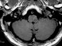
Figure 1
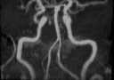
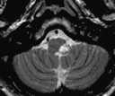
Figure 2
Figure 1 T1WI shows a hypointense focus in the left lateral medulla. A high signal crescent rim is noted in the wall of the left vertebral artery.Figure 2 The medullary lesion shows high signal intensity on T2WI.Figure 3 MRA demonstrates luminal narrowing in the left vertebral artery.
| ||||
HISTORY |
PREVIOUS MTHS |
|||
HOME |
CURRENT MTH |
|||