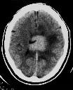
Figure 1
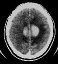
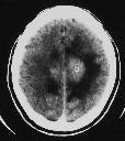
Figure 2
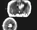
Figure 1 NECT brain shows a hyperdense mass in the corpus callosum and adjacent peritumour oedema.Figure 2 NECT brain shows the mass to be seperated from the falx.Figure 3 CECT brain demontrates the strong and homogenous contrast enhancement of the mass.Figure 4 Coronal reformat image of the brain clearly shows the mass to be in the corpus callosum. Biopsy revealed lymphoma of brain.
| ||||
HISTORY |
PREVIOUS MTHS |
|||
HOME |
CURRENT MTH |
|||