CLINICAL HISTORY:
A newborn baby girl found to have a large abdominal mass in antenatal US.
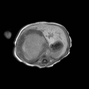
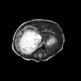
CT and MRI was performed.
DIAGNOSIS:
Infantile Hemangioendothelioma
DISCUSSION:
Infantile hemangioendothelioma is the commonest benign hepatic tumour of infancy. 90% diagnosed in the first 6 months of life. Most manifest as an asymptomatic abdominal mass. Possible but uncommon complications include CHF or tumour rupture.
In US, infantile hemangioendothelioma is commonly presented as a well demarcated hypoechoic mass. Typical hyperechoic appearance of adult hemangioma is uncommon Enlarged hepatic vessels are usually seen due to high flow nature of the lesion.
In CT, the tumour is usually hypodense to normal liver, with enhancement pattern similar to adult hemangioma, that is peripheral nodular enhancement in arterial phase, and progressive centripetal fill-in at later phases. Speckled calcifications are seen in 50% of the tumours, and large focal tumour will have central necrosis or hemorrhage.
For MRI, the tumour is hypointense to normal liver in T1WI, and markedly T2 hyperintense due to vascular nature. Flow voids are also common adjacent to tumour.
Differentials include hepatoblastoma or metastatic lesions. In the former, it is usually more solid and less vascular. The tumour is uncommon in newborn and AFP is usually elevated. For the latter, they are commonly multifocal, and further workup should be done to identify the primary site.
Biopsy of the tumour should be avoided due to its highly vascular nature.
|