|
|
|
|
|
|
|
|
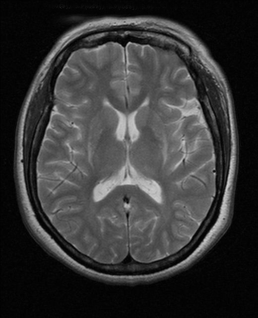
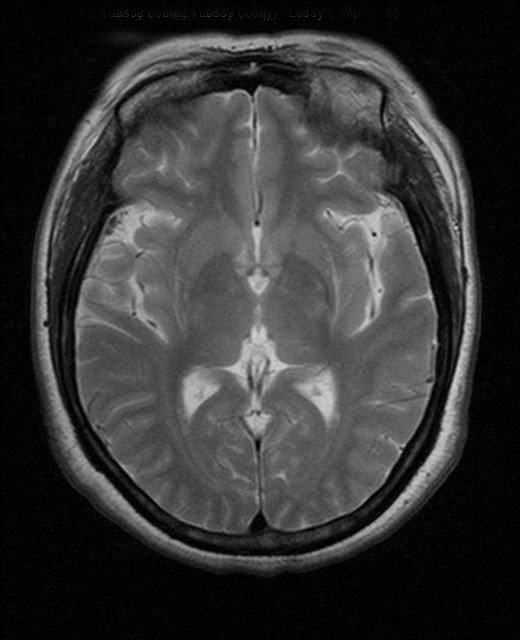
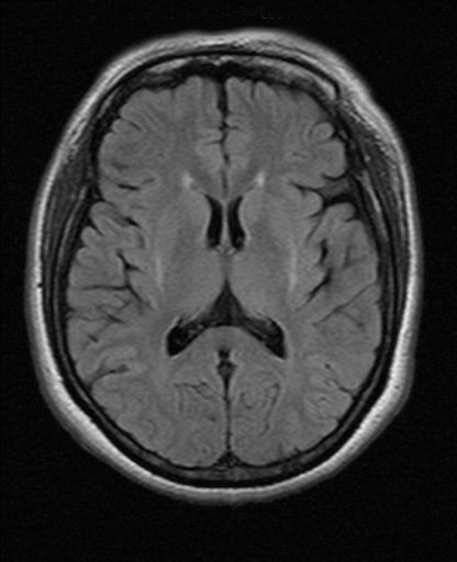
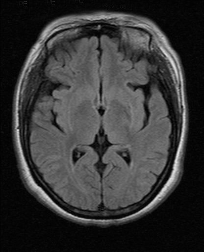
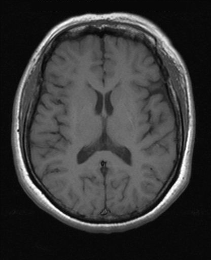
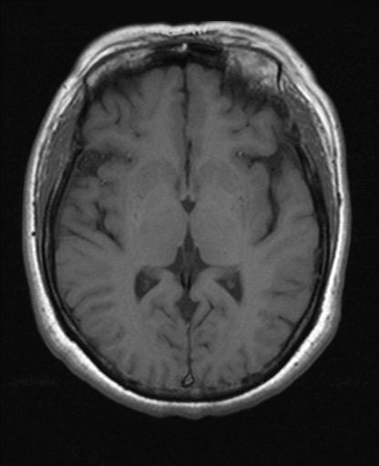
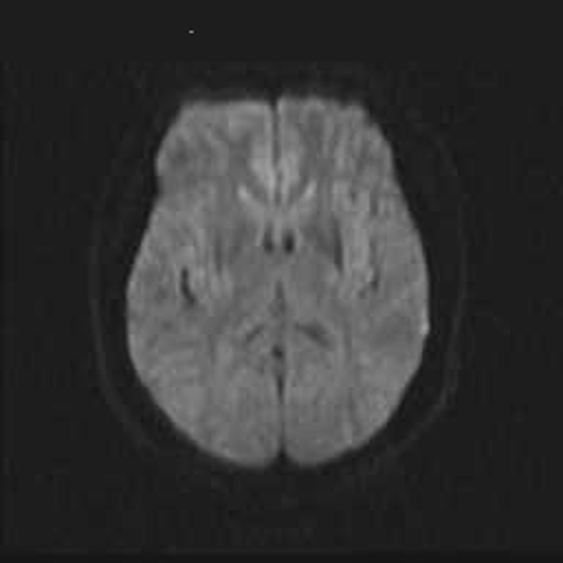

CLINICAL HISTORY: MRI brain was performed to rule-out organic cause. |
||
PREVIOUS CASES |
||
HOME |
COMMENTS |
|
CASE OF THE MONTH |
|
|
|
|||||||||||||||||
|---|---|---|---|---|---|---|---|---|---|---|---|---|---|---|---|---|---|---|
Click on thumbnail for full-sized image
Disclaimer: The material used in this Web page is for educational purpose only.