|
|
|
|
|
|
|
|
|
|
|
|


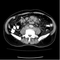
CLINICAL HISTORY: DIAGNOSIS: DISCUSSION: Mesenteric panniculitis is twice as common in males than females. The typical presentation is one of chronic abdominal pain, fever and weight loss. Alternatively, the patient may be asymptomatic and the finding is seen incidentally on CT. CT features of mesenteric panniculitis vary depending on the predominant tissue. It could be increased density of mesenteric fat, mesenteric vessel encased by increased density fat, scattered soft tissue mesenteric nodules, “fat ring sign” i.e. preservation of fat around vessels and “pseudocapsule” sign i.e. peripheral band of soft tissue attenuation that limits normal mesentery from inflammatory process. In some patients (reports range from 1% to 70%), the presence of mesenteric panniculitis is a sign of malignancy elsewhere. These malignancies include lymphoma, breast cancer, lung cancer, melanoma and colon cancer. As in this case, diffuse large B cell lymphoma is present. In the absence of malignancy, most patients with mesenteric panniculitis have a favourable prognosis and may be managed conservatively. In our case, the patient is treated with cycles of chemotherapy. |
||
PREVIOUS CASES |
||
HOME |
COMMENTS |
|