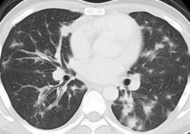
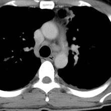
CASE OF THE MONTH

2011

June


CXR at presentation


CXR after 2 months

CLINICAL HISTORY:
A 32-year-old man with a newly diagnosed systemic disease complained of shortness of breath. Serial chest X rays and a set of CT thorax were performed.

DIAGNOSIS:
Kaposi Sarcoma

DISCUSSION:
CXR showed bilateral linear opacities and patchy areas of consolidation, which has a bilateral perihilar distribution of middle and lower zone predominance. There is interval deterioration on follow up CXR after 2 months. CT showed multiple flame-shaped lesions in both lungs. There are bronchial wall and interlobular septal thickening. Mediastinal lymph nodes are enlarged. These features are characteristic of Kaposi sarcoma in patients with HIV infection.

Kaposi sarcoma (KS) is the most common AIDS-related neoplasm. It predominantly affect homo- or bisexual men. There is an association with human herpesvirus 8. Histological findings would show spindle cell proliferation with extravasated red cells within stromal clefts. Skin is the most frequently affected site. Pulmonary involvement is a late complication of AIDS.

KS show a peribronchvascular distribution.  Imaging findings include peribronchial cuffing, septal line thickening, perihilar coalescent consolidation, flame-shaped pulmonary nodules, pleural effusion and lymphadenopathy.

PREVIOUS CASES

HOME

COMMENTS

Click on thumbnail for full-sized image


Disclaimer: The material used in this Web page is for educational purpose only.