|
|
|
|
|
|
|


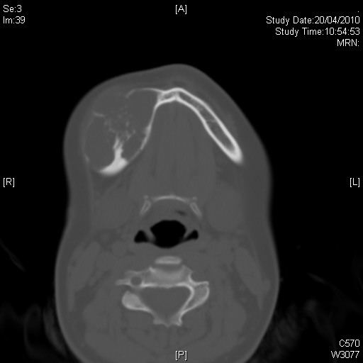


CLINICAL HISTORY: DIAGNOSIS: Ameloblastoma DISCUSSION: Ameloblastoma is a benign, but locally aggressive odontogenic lesion. It commonly arises from the ramus and posterior body of the mandible. Typically, it is a mixed cystic and solid lesion with soap-bubble appearance, and its associated with the crown of an impacted or unerupted tooth, resorption of the adjacent root of a tooth is also a common finding. However, they can vary in the radiological appearance, some of them are very well-defined, unilocular, well-corticated, lucent lesions that are often associated with the crowns of impacted or unerupted teeth, as a result, such ameloblastomas are indistinguishable from odontogenic keratocysts and dentigerous cyst radiologicaly. Other ameloblastomas are multilocular with internal septa and a honey-comb or soap bubble appearance and are often similar in appearance to odontogenic keratocysts. In most of the cases, radiological features are unable to make a definitive diagnosis, so these lesions should be surgically removed and examined microscopically to accurately establish the diagnosis.
|
||
PREVIOUS CASES |
||
HOME |
COMMENTS |
|