CLINICAL HISTORY:
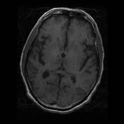
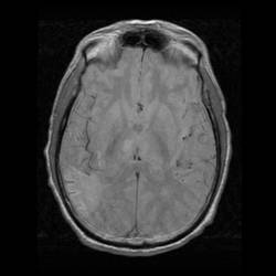
A 60 year old man with history of atrial fibrillation and chronic renal failure presented with generalized limb weakness and decreased general condition with GCS 7/15. Plain CT brain scan and subsequently MRI brain scan were done during hospital stay.
DIAGNOSIS :
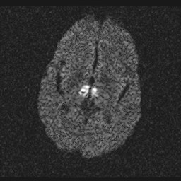
Percheron artery occlusion
DISCUSSION :
The thalami receive their blood supply from both the anterior and posterior circulation. The posterior communicating arteries of the anterior circulation give thalamoperforating arteries and supply the anteroinferior aspects of thalami. In the posterior circulation, segment P1 and P2 of the posterior cerebral arteries supply the medial, lateral and superior aspects of the thalami.
A thrombus at the tip of basilar artery will occlude blood flow in bilateral posterior cerebral artery can give rise to bilateral thalamic infarct and possibly infarct in brainstem, inferior surface of temporal and occipital lobes.
Percheron artery is a variant of the posterior arterial circulation of the brain. It is a common trunk arising from one of the P1 segments providing bilateral distribution. Occlusion of Percheron artery will lead to infarcts in bilateral medial thalami and brain stem.
The above two differentiate diagnosis could be differentiated by angiogram.
Venous thrombosis of the internal cerebral vein or vein of Galen can also give rise to ischemia in posterior fossa. However, it is differentiate from the above by extensive edema and occasional hemorrhagic change. |