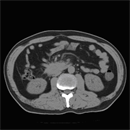
Fig.1
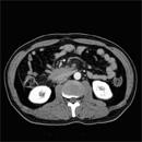
Fig.2
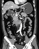
Fig.3
Diagnosis : Desmoid( Retroperitoneal Desmoid).Findings :Figures 1 and 2 show slightly enhancing irregular infiltrative mass at root of mesentery adjacent to third part of duodenum. No calcification is seen inside the mass and it is not associated with mesenteric adenopathy. Figure 3 is coronal 3D reformat image showing the mass at roof of mesentery separated from head of pancreas. The stomach and duodenum are also distended proximal to the mass. |
||
DIAGNOSIS : |
Pulmonary varix t |
|
PREVIOUS CASES |
||
HOME |
COMMENTS |
|