
Figure 1
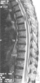
Figure 2
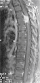
Figure 3
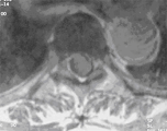
Figure 4
Figure 1: T1 weighted sagittal image shows an isointense lesion expanding the cord at T5.Figure 2: T2 weighted sagittal image shows that the lesion at T5 is isointense.Figure 3: T1 weighted postcontraast image shows that the lesion is intensely enhanced and appear hyperintense. There is "dural tail" sign.Figure 4: T1 weighted postcontrst axial image at T5 level suggests that the lesion is intradural extramedullary in location. |
||
DIAGNOSIS : |
Meningioma |
|
PREVIOUS CASES |
||
HOME |
COMMENTS |
|