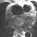
Figure 1
Figure 3
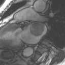
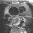
Figure 2
Figure 4
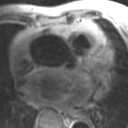
Figure 1 & 2 Axial post-contrast scan of calf showing abnormal filling defects in the deep veins of both lower limbs.Figure 3 Coronal post-contrast axial scan of calf showing thrombus in deep veins of both lower limbs.Figure 4 axial scan of pelvis showing abnormal soft tissue mass behind uterus. During operation the mass is confirmed to be carcinoma of ovary.
| ||||
HISTORY |
PREVIOUS MTHS |
|||
HOME |
CURRENT MTH |
|||