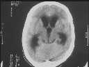
Figure 1
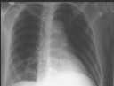
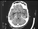
Figure 2
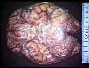
Figure 1 CT showed hydrocephalus.Figure 2 CECT basal enhancement not prominent.Figure 3 CXR showed consolidation in posterior basal segment of left lower lobe.Figure 4 Post-mortem showed TB meningitis with endarteritis and thrombosis, abscess in left lower lobe and miliary TB in lung.
| ||||
HISTORY |
PREVIOUS MTHS |
|||
HOME |
CURRENT MTH |
|||